Introduction:
Recently, research groups of Prof. Tao Li’s and N.A. Shining Zhu’s of College of Engineering and Applied Science, working with Dingping Cai’s research group of Academia Sinica, made crucial progress in imaging with aplanatic metalens. They developed nonmotion focal length tuning and spectral tomography based on the large chromatic dispersion of metalens. They also demonstrated microscopic spectral tomography of biocells, which shows the great potential of the applications of aplanatic metalens in highly integrated and stable imaging systems. The manuscript titled as Spectral Tomographic Imaging with Aplanatic Metalensis published on the top international optical journal Light: Science & Applications 8, 99 (2019)(IF=14.0). Prof. Tao Li of College of Engineering and Applied Science is the corresponding author of the paper. Chen Chen, a Grade 16 bachelor-straight-to-doctorate student, is the first author. Another Grade 16 bachelor-straight-to-doctorate student Wange Song along with Jiawen Chen of Academia Sinica also contribute a lot.

Figure 1 Schematic of microscopic spectral tomographic imaging of biocells by metalens
Background:
Metasurfaces, capable of flexibly manipulating the amplitude, phase, and polarization of light by subwavelength units, are the most attractive flat optical devices at the moment. The metalens has captured significant attention as one of the flat optical device with great potential. Great efforts have been made towards its practical applications, such as efficiency improvement, chromatic aberration correction, and image correction. Over the past years, Shining Zhu, Tao Li, and Shuming Wang’ research groups of Nanjing University cooperated with Dingping Cai’s of Taiwan and developed broadband achromatic optical metasurface devices, as well as broadband achromatic metalens both in the visible and near-infrared region. Related works are published on Nature Communications 8, 187 (2017), Nature Nanotechnology 13, 227 (2018), Nature Nanotechnology 14, 227 (2019). However, due to the limited range of phase compensation, restriction is imposed on the size and numerical aperture of those achromatic metalens, which hinders its application badly. In this work, Tao Li and Chen Chen in turn utilized the large chromaticity of metalens as a tuning dimension to access tunable functionalities. Moreover, aplanatic design was introduced to guarantee a high longitudinal resolution to obtain high-resolution spectral tomographic imaging. Conventional tomography is usually implemented by mechanical scanning which brings complexity and instability. The metalens with large chromatic dispersion introduced by this work requires no phase compensation and thus the size of device is unlimited. In the meantime, the nonmotion spectral imaging breaks the limitation of the depth of filed of conventional lens and greatly improves integration and stability.
Creative research:
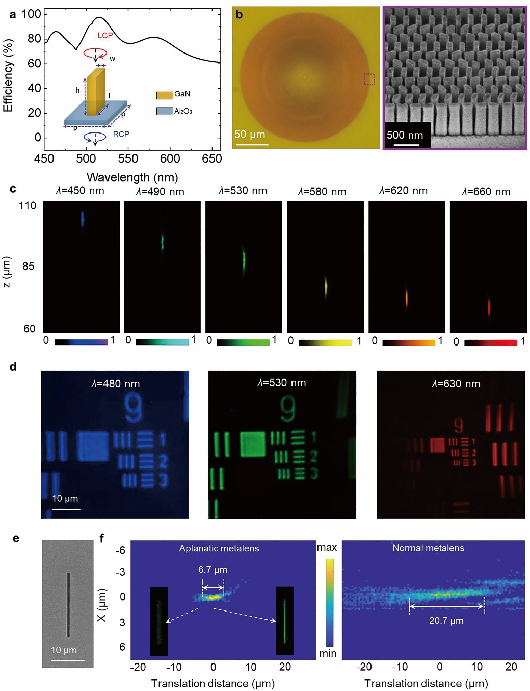
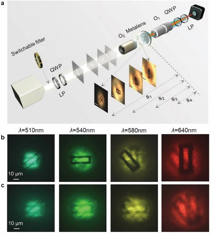
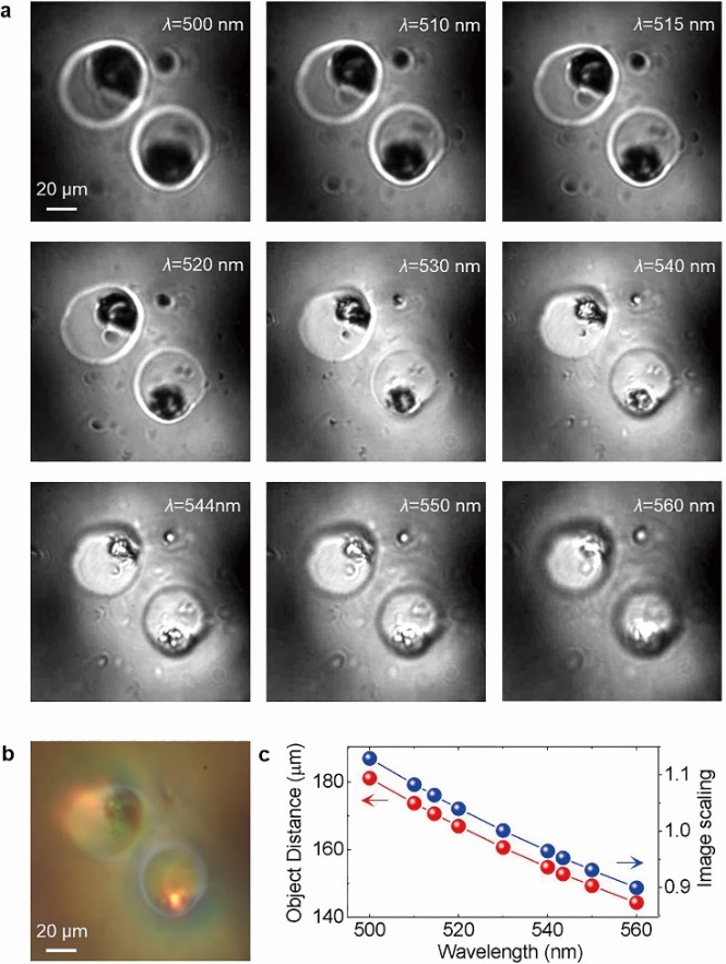
In this work, the research group demonstrated a brand new nonmotion spectral microscopic tomography technique, designed and constructed the aplanatic GaN metalens, with which high-resolution spectral tomographic images of biocells were obtained(Fig. 1). This work first designed the aplanatic phase distribution of the metalens with high numerical aperture, which significantly improved the transverse / longitudinal resolution and showed considerable bandwidth(Fig. 2). Next, the research group designed and processed the high efficiency aplanatic GaN metalens in the visible spectrum(450 nm-660 nm) based on berry phase method and characterized the diffraction dispersion, transverse and longitudinal resolution of the metalens in detail(Fig. 3). It could be observed that aplanatic metalens showed obvious advantages compared with conventional metalens in the above aspects. Then, an object with multiple layers were used to verify the tomographic imaging of metalens. When the image distance was fixed, utilizing the fact that the focal length of the metalens varies with wavelength, the image and depth information of objects with different depths were measured, which confirmed the function of tunable focal lengths and tomography of the aplanatic metalens(Fig. 4). Based on the above characterization the researchers took a step further and demonstrated microscopic spectral tomographic images of frog egg cells. According to the focused images of different wavelengths, the sizes of nucleus and cell membrane can be estimated(Fig. 5). At last, this work compared metalens with conventional diffractive lens at length and revealed that the unique polarization manipulation of metalens could greatly improve the signal-to-noise ratio of tomographic imaging. This work creatively utilizes the chromaticity of metalens and demonstrates a high-resolution and high signal-to-noise ratio microscopic tomographic imaging technique, which breaks the restriction on the size of numerical aperture and lens, showing great potential for applications in highly integrated and stable imaging systems.
This research is supported by the National Key R&D Program of China, the National Natural Science Foundation of China, Mountain-Climbing Talents Program of Nanjing University, and many other programs.
Link of the paper:https://www.nature.com/articles/s41377-019-0208-0
Quick view of the images:

Figure 2 Phase design of the aplanatic metalens.(a)Phase profiles;(b)Spherical aberrationas a function of the numerical aperture;(c)Normalized spherical aberration,showing the broadband performance;(d) Comparison of simulation results of the point-source imaging.

Figure 3 Characterization of aplanatic metalens.(a)Calculated conversion efficiencyof GaN unit;
(b)Optical and SEM images of the aplanatic metalens with NA = 0.78;(c)Dispersion;
(d)Characterization of transverse resolution in the visible region(at least 775nm);
(e)Slit for longitudinal resolution measurement;(f)Comparison of longitudinal resolution.

Figure 4 Spectral tomography of object with multiple layers.(a)Schematics of the imaging setup;(b) Tomographic image of the aplanatic metalens;(c) Tomographic image of the normal metalens.

Figure 5 Microscopic tomographic images of frog egg cells.(a) Microscopic tomographic images at different wavelengths;(b)Direct white-light image;(c)The derived layer positions and imaging scalings at different wavelengths.
(Submitted by Chen Chen)

